


Rapid advances in the field of endovascular neurosurgery and the development of minimally invasive techniques have resulted in a great expansion of potential therapeutic applications. Two possible applications are
- revascularization leading to reopening of blood vessels
- embolization leading to occlusion of blood vessels
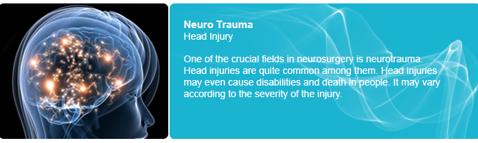
ACUTE ISCHEMIC STROKE:
There has been improvement in outcome of stroke after the advent of intravenous (IV) recombinant tissue plasminogen activator (rtPA) for the treatment of acute ischemic stroke. The intra arterial (IA) approach is believed to have the advantage of delivering thrombolytic agents to the thrombus in high concentrations locally. Theoretically, this allows lower systemic dose and assumed lower hemorrhagic complication rates. It also allows the simultaneous use of mechanical disruption to facilitate thrombolysis.
The potential time delay required to obtain the cerebral angiography and place the microcatheter in proper position to administer the thrombolytic agent is a disadvantage of this modality, hence the idea of combing IV thrombolysis with the IA approach. The Emergency Management of Stroke (EMS) Bridging trial was a randomized, double-blind, placebo-control study which demonstrated a higher recanalization rate (53%) in the combined IV/IA alteplase treatment group versus the IA alteplase group (28%). This suggested the feasibility of the approach. Bridging is favoured in a subset of patients who are expected to have a limited response to IV treatment such as those with severe neurological deficits, presentation between 3 and 6 hrs of symptom onset, history of major surgery with in the last 14 days, and occlusion of major cervical or intracranial vessels with a high National Institute of Health Stroke Scale (NIHSS) score.
The mechanical devices used for treatment of stroke can be classified into two main groups.
- mechanical disruption devices such as snares, balloon mounted stents, angiojets , neurojets etc.
- mechanical clot retrieval devices such as the Merci retrieval system, penumbra aspiration device.
CAROTID STENOSIS:
Accumulating evidence has shown th ebenfit of carotid endarterectomy in reducing the risk of stroke and death in patients with moderate to severe symptomatic (greater than 50%) or aymtomatic (greater than or equal to 60%) carotid stenosis.
Perioperative complication rates in high risk patients have exceeded the potential benefits in general practice and hence high risk patients were omitted from the study population. This is especially true in patients with unfavorable clinical and anatomical characteristics.
Carotid angioplasty with stent placement (CAS) was initiated as an alternative treatment for revascularization of carotid artery stenosis in high risk surgical candidates. More recently, several non randomized studies and randomized comparisons with carotid endarterectomy have increased the popularity of CAS. These studies have focused on the safety and effectiveness of CAS. With improving devices and techniques, CAS has become safer than and at least as effective as surgical treatment. Studies have strongly advocated the use of a distal protection device in high risk surgical patients.
It is well known that patients undergoing CAS have small embolic showers occurring frequently during the procedure that can persist with increased frequency compared with baseline for hours or days after the angioplasty. This has been shown using intra procedural trans cranial doppler imaging (TCD). It is also known that these microemboli are composed of thrombotic and plaque substances. MRI had suggested that about 15% of patients developed new ischemic lesions based on diffusion related abnormalities in the brain after this procedure. A similar study suggests that almost half of these patients remain symptomatic. From previous data on carotid endarterectomy, it appears that the rate of new brain lesions is higher in CAS than the rate reported for endarterectomy at 5 %. This underlies the importance of using a distal protection device to prevent the microemboli from being released into cerebral circulation during carotid angioplasty and stenting.
INTRACRANIAL AND VERTEBRAL ARTERY STENOSIS:
Intracranial stenosis is responsible for 8-10% of all ischemic strokes. There is an astoundingly high yearly rate of recurrent strokes in patients with intracranial stenosis that has been estimated at approximately 8-12%. For those patients not responding to antithrombotic treatment, the rate of recurrent ischemic stroke can be even higher and has been estimated at 52%. Patients with symptomatic vertebro basilar stenosis had a low stroke free survival rate of 76% at 12 months and 48% at 5 years.
Developments such as intracranial balloons and stents specific for atherosclerotic lesions with greater radial force such as the Wingspan angioplasty and stent system could also further advance intracranial angioplasty and stenting. It is expected that in the future a combination of mechanical protection devices and better designed stent delivery systems, as well as drug eluting stents, should help to reduce perioperative and post operative complication rates.
SUB ARACHNOID HEMORRHAGE DUE TO INTRACRANIAL ANEURYSMS:
Subarachnoid hemorrhage (SAH) is a common and often devastating occurrence. Intracranial aneurysms constitute an important health problem worldwide, affecting about 2% of the population.
The first reported endovascular attempt at approaching an aneurysm was reported in 1941, when Werner and colleagues inserted silver wires via transorbital approach to prevent rupture of a susceptible wall of an aneurysm from the stress of pulsatile blood. Subsequently, placement of particles and other agents including platinum coils was attempted. By the late 1980s, platinum coils have been introduced. These were attached to a steel delivery wire system that could be delivered through a micro catheter to aneurysmal sac. Subsequently an electrical current applied to the wire attached to the coil was used to detach the stainless steel wire from the platinum coil. The protection provided by the coil inside the aneurysm was thought to be due to immediate hemodynamic changes and redirection of the blood flow through the proper vessel. An eventual thrombus formed within the aneurysm that protects the osteal neck of the aneurysm and eventually fills the inner cavity with connective tissue.
The endovascular coils are placed in the aneurysmal sac either with the assistance of a balloon, stent, another micro catheter itself. Better coil designs have improved the ability to obliterate the aneurysmal sacs.
Initially, endovascular treatment was reserved for patients who were regarded as poor candidates for surgical clipping. It included patients who were elderly, those wit poor Hunt- Hess grades IV or V, those whose aneurysms are located in the posterior circulation or were located in cavernous segment of internal carotid artery and had active cerebro vasospasm. The larger randomized multicenter phase III trial (International Subarachnoid Aneurysm Trial, ISAT), compared the safety and efficacy of embolization with neurosurgical clipping. ISAT enrolled 2143 patients with ruptured aneurysms who were considered suitable for either treatment. The neurosurgical clipping group had 1070 patients and the group receiving endovascular treatment by detachable platinum coils had 1073 patients. The clinical outcomes were assessed at 2 months and at 1 year with interim ascertainment of re bleeds and death. Of the 801 patients allocated to endovascular treatment, 190(23.7%) became dependent or died at 1 year compared with 243(30.6%) of 793 allocated to neurosurgical treatment. The risk of re bleeding from ruptured aneurysms after 1 year was 2 per 1276 and 0 per 1081 patients allocated to endovascular and neurosurgical treatment respectively. It was concluded that the outcomes in terms of survival free disability at one year was significantly better with endovascular coiling.
The recently published guidelines, from the Stroke Journal (Feb 2009) recommends For patients with ruptured aneurysms judged by an experienced team of cerebrovascular surgeons and endovascular practitioners to be technically amenable to both endovascular coiling and neurosurgical clipping, endovascular coiling can be beneficial (Class I, Level of Evidence B).
Considerable advances have been made in the ability to use coils for endovascular treatment of intracranial aneurysms in situations that might have appeared unsuitable a few years ago. The new designs of coils, including the three and two dimensional configurations, have improved results. Focusing on the anatomy of the aneurysm and on the neck and dome ratio, as well as its packing with coils in a step wise manner, has led to remarkable success in coiling of aneurysms previously considered to be difficult.
ARTERIOVENOUS MALFORMATIONS:
CNS arteriovenous malformations (AVMs) occur in approximately 0.15% of the population. Of these, 90% are supratentorial in location. There is a rate of approximately 4% hemorrhagic conversion per year for cerebral AVMs. They carry a combined morbidity and mortality rate of 2.7% per year. AVMs can cause headache, seizures and ischemia. The treatment of AVMs is challenging and multifaceted. They pose a unique problem and often require a combination of therapeutic modalities. Embolization of cerebral AVMs on its own is curative in fewer than one quarter of lesions. Embolization has been used for the following purpose.
- adjunct to surgery
- eduction of size before radiation
- palliation
- embolization alone for cure.
Embolization is performed using a micro catheter placed in a distal arterial feeder that supplies the AVM nidus exclusively. The nidus of the AVM is the focus of embolization procedure. Any abnormal branches are excluded on the basis of assessment of vessel architecture and at times physiological testing by injection of short acting amobarbitol sodium and lidocaine through a micro catheter. There are different embolic materials available. Solid embolic agents consist of poly vinyl alcohol particles, fibers, micro coils and micro balloons. Liquid embolic agents consist of cyanoacrylate monomers and polymer solutions such as ethyl vinyl alcohol polymer.
The usual goal of pre surgical embolization is to decrease the nidus size and prevent perioperative blood loss associated with AVM resection. Hence this enhances the safety and ease of operative intervention. Embolized AVM vessels can also serve as a road map during surgical intervention. Because AVM surgery carries a certain amount of risk, AVMs that would significantly benefit from embolization before surgery should undergo this treatment. For example, a small AVM in the right frontal lobe with easily accessible arterial feeders may not warrant embolization before surgery. Also, radiosurgery has emerged as a relatively safe way to treat small AVMs. Two indirect comparisons have suggested that embolization before surgery can shorten operative time reducing intra operative blood loss and can improve neurological outcomes in patients with complex AVMs such that they are comparable to those with less complex lesions.
TRAUMATIC NEUROVASCULAR INJURY:
Transcatheter occlusion of traumatically injured vessels to control life threatening bleeding represents some of the earliest endovascular procedures. Penetrating injury to the arteries of the head and neck may lead to dissection and pseudoaneurysm formation with the dreaded consequence of high pressure arterial hemorrhage. Endovascular stenting of dissected intimal flaps aids in tacking down the affected vessel wall and preventing down stream dissection. This has been well described for injuries to the carotid, vertebral and subclavian arteries.
HEAD AND NECK TUMORS:
The indications for CNS tumor embolization are
- achieving control of surgically inaccessible arterial feeders
- decreasing surgical morbidity by reducing operative blood loss
- shortening operative procedural time
- improving the likelihood of complete surgical resection
- decreasing the risk of damage to the adjacent normal tissue
- relieving intractable pain
- decreasing tumor recurrence
- allowing better visualization of the surgical field
- reducing surgical complications.
FUTURE DIRECTIONS:
Endovascular therapy for acute and chronic cerebrovascular diseases and interventional neuroimaging is evolving at a rapid pace. The safety and efficacy of endovascular procedures is expected to improve wit new technologies. Techniques for preventing restenosis with the advent of drug eluting stents have already advanced at a rapid pace. Further developments in stent design would help to address the issue of restenosis in smaller diameter blood vessels. Further designs of devices for aneurysm obliteration and newer intracranial supportive stents might help for a more durable approach to aneurysm coiling. Biopolymer coated coils heave been suggested to increase clot maturation and enhance intra aneurysmal fibrosis and thereby potentially decrease recanalization rates. A combination of endovascular surgery in AVM treatment with rapidly advancing radiosurgery might make it possible for most AVMs to be treated in a minimally invasive manner in the near future. Improvement in AVM embolization may be possible with the development of liquid embolic agents.
Endovascular procedures are rapidly expanding as treatment options for cerebrovascular diseases and neoplasms and are becoming less invasive but more effective.. It is essential to understand the scientific basis of treatment rationale based on advancing neuro imaging techniques to better serve our patients.